The Radio Vision Graphy (RVG) imaging system1 commonly
used in dentistry to take intraoral periapical radiographs features the latest
innovations in digital radiography, delivering the highest image resolution
(> 20 LP/mm). RVG consists of a sensor, monitor, and microcomputer
components
What is a periapical
radiographic image?
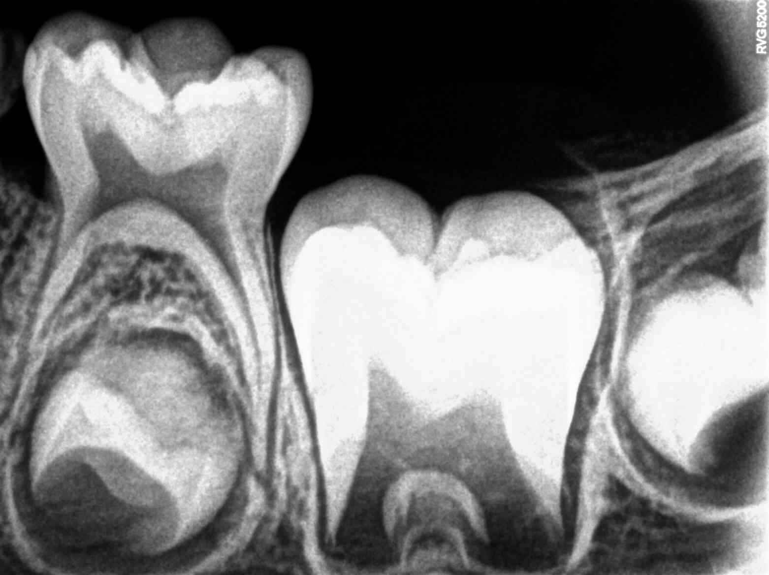
A
periapical x-ray is one that captures the whole tooth. It shows everything from
the crown (chewing surface) to the root (below the gum line). Each periapical
x-ray shows a small section of your upper or lower teeth. These x-rays are
often used to detect any unusual changes in the root and surrounding bone
structures.
What are the
limitations of periapical radiography?
The
periapical radiography is nowadays the main radiographic investigations used
but presents some limits as 3D anatomic alteration, geometric
compression, and possible anatomical structures overlapping that can
obscure the area of interest.